
Coxsackieviruses are a subset of the genus Enterovirus and belong to the Picornaviridae family. For the first time, the Coxsackie viruses were isolated by G. Sikels and G. Daldorf in 1948. Coxsackieviruses cause a variety of acute infectious diseases – from summer flu and herpangina to viral meningitis, vesicular stomatitis, limb exanthema, meningitis and pneumonia in newborns. The viruses are divided into two groups – A (CVA) and B (CVB), which are based on their effects on experimental mice (coxsackievirus A leads to muscle damage, paralysis and death, coxsackievirus B leads to organ damage, but less severe results.) In humans, group A coxsiviruses tend to infect the skin and mucous membranes, causing herpagin, acute hemorrhagic conjunctivitis, and foot-and-mouth disease. Group B – the heart, pleura, pancreas and liver, causing myocarditis, pericarditis and hepatitis. Both groups A and B can cause non-specific febrile conditions, rashes, upper respiratory tract diseases and aseptic meningitis.
Morphology and cultural properties
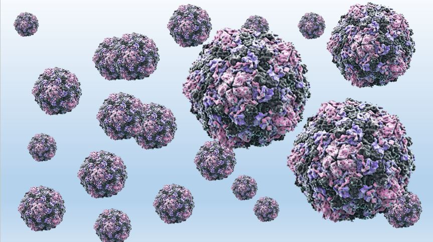
Coxsackieviruses have the RNA genome, their size is about 28-30 nm, and their shape is ixohedral. They are propagated in cell cultures by human amnion and kidney. They cause cell cytolysis (destruction) and can be demonstrated by a viral neutralizing reaction. At least 23 serotypes (1-22, 24) of group A and 6 serotypes (1-6) of group B. are known.
Epidemiology
Coxsackie virus infections are widespread in the world. They can be isolated year-round in tropical climates, with decreasing disease incidence and seasonality in areas of greater latitude. Outbreaks of Coxsackie B virus infection occur mainly in spring and summer, when enteroviruses are most prevalent.
Pathogenesis and clinical presentation
Coxsackieviruses are mainly transmitted by the faecal-oral and airborne droplets. Viruses initially reproduce in the upper respiratory tract and the epithelium of the digestive tract. They are found in the respiratory tract up to 3 weeks after the initial infection and in the stool up to 8 weeks. Viruses have been found to replicate in submucosal lymphoid tissue and spread to the reticuloendothelial system. Further distribution in the target organs (throat, heart, liver, skin) occurs after secondary viremia. More than 90% of coxsackievirus infections are asymptomatic or cause non-specific febrile conditions. In newborns, they are the most common cause of fever in the summer and fall. In 13% of infants with fever, enterovirus infection is proven within the first month of life.
Coxsackie viruses can cause:
- Herpangina
It is caused by the coxakivirus group A, serotype 16. They are characterized by acute onset fever up to 40 degrees, fatigue, sore throat, hyperemia and vesicular rash of the pharynx. It lasts about 2-3 days.
Vesicular stomatitis with exanthema
It flows with a vesicular rash on the mucous membranes of the mouth and skin of the palms, wrists, fingers and soles. It mainly affects children aged 1 to 8 years.
Aseptic meningitis
Patients with aseptic meningitis may have:
- rapid or gradual onset of fever and chills;
- nausea and vomiting;
- headache;
- neck rigidity;
- sensitivity to light.
Infants less than 3 months of age have the highest incidence of clinically recognized aseptic meningitis, with only fever, irritability and anorexia. No long-term neurological deficits have been observed in infants with aseptic meningitis. Older adults may have a longer period of fever and headache than babies and children.
Neurological diseases
Rarely, coxakiviruses are implicated in additional neurological diseases such as sporadic cases of low motor paralysis, which is very similar to poliovirus infection.
Mortality due to coxsackievirus infection is rare. Newborns and immunocompromised individuals are at highest risk for complications.
Microbiological diagnostics
Investigating:
- throat secretion;
- feces;
- vesicular fluid;
- blood;
- liquor.
Preparation of the preparations consists of suspension in Hanks solution, centrifugation and antibiotic treatment (to remove the accompanying microflora). Inoculation of white mice, cell culture infestation, and serological diagnosis are the best methos of Virus isolation.